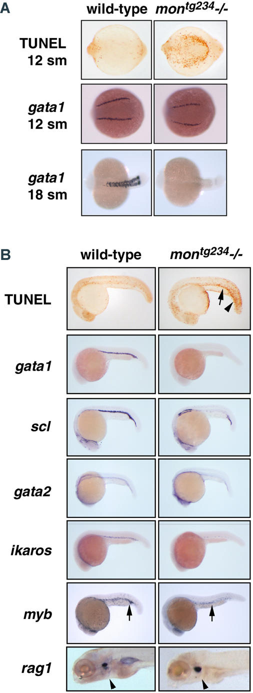
Figure 1. Zebrafish mon Mutants Have Severe Defects in Primitive Hematopoiesis.
(A) Whole-mount TUNEL assays reveal that ventral-posterior mesodermal cells undergo apoptosis in homozygous montg234 mutant embryos. Whole-mount in situ hybridization of gata1 detected at the 12- and 18-somite stage in genotyped embryos. Posterior views, anterior to the left.
(B) Extensive apoptosis is visible in the trunk and tail (arrowhead) and also in hematopoietic cells of the embryonic blood island at 22 h of development (arrow). Whole-mount in situ hybridization at 22 hpf including scl, gata2, gata1, ikaros, and myb in montg234 mutants. Expression of myb is greatly reduced in the blood islands because of a loss of erythroid cells, but embryonic macrophages are still present (arrows). The expression of rag1 in thymic T-cells appears normal in mon mutants at 5 d postfertilization (arrow heads). Lateral views of 22 hpf and 5-d-old embryos.

