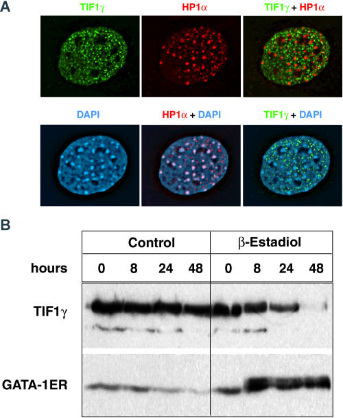
Figure 6. Mammalian Tif1γ Protein Localizes to Nuclear Bodies Distinct from Heterochromatin.
(A) Deconvolved immunofluorescence images of a mouse embryonic fibroblast cell nucleus stained with an anti-Tif1γ antibody and stained with a monoclonal antibody directed against HP1α. This is also compared to DAPI staining. The merged images of the nucleus show that Tif1γ does not colocalize with the HP1α or DAPI staining of heterochromatin while HP1α and DAPI staining overlap.
(B) G1ER mouse erythroleukemia cells express high levels of Tif1γ protein as detected by Western blot analysis. Expression of Tif1γ decreases during Gata1-dependent erythroid maturation induced by β-estradiol treatment to induce a Gata1–ER fusion protein.

