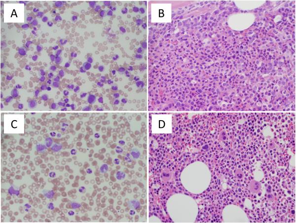
Figure 1. Blood and bone marrow morphology.
(A) Peripheral blood smear at CML diagnosis demonstrating marked leukocytosis with granulocytic left shift and decreased platelets. (B) Bone marrow biopsy at CML diagnosis shows hypercellularity with granulocytic hyperplasia. (C) Peripheral blood smear on day 92 of imatinib therapy showing leukocytosis with monocytosis and (D) corresponding bone marrow biopsy showing hypercellular bone marrow with occasional hypolobated megakaryocytes.

