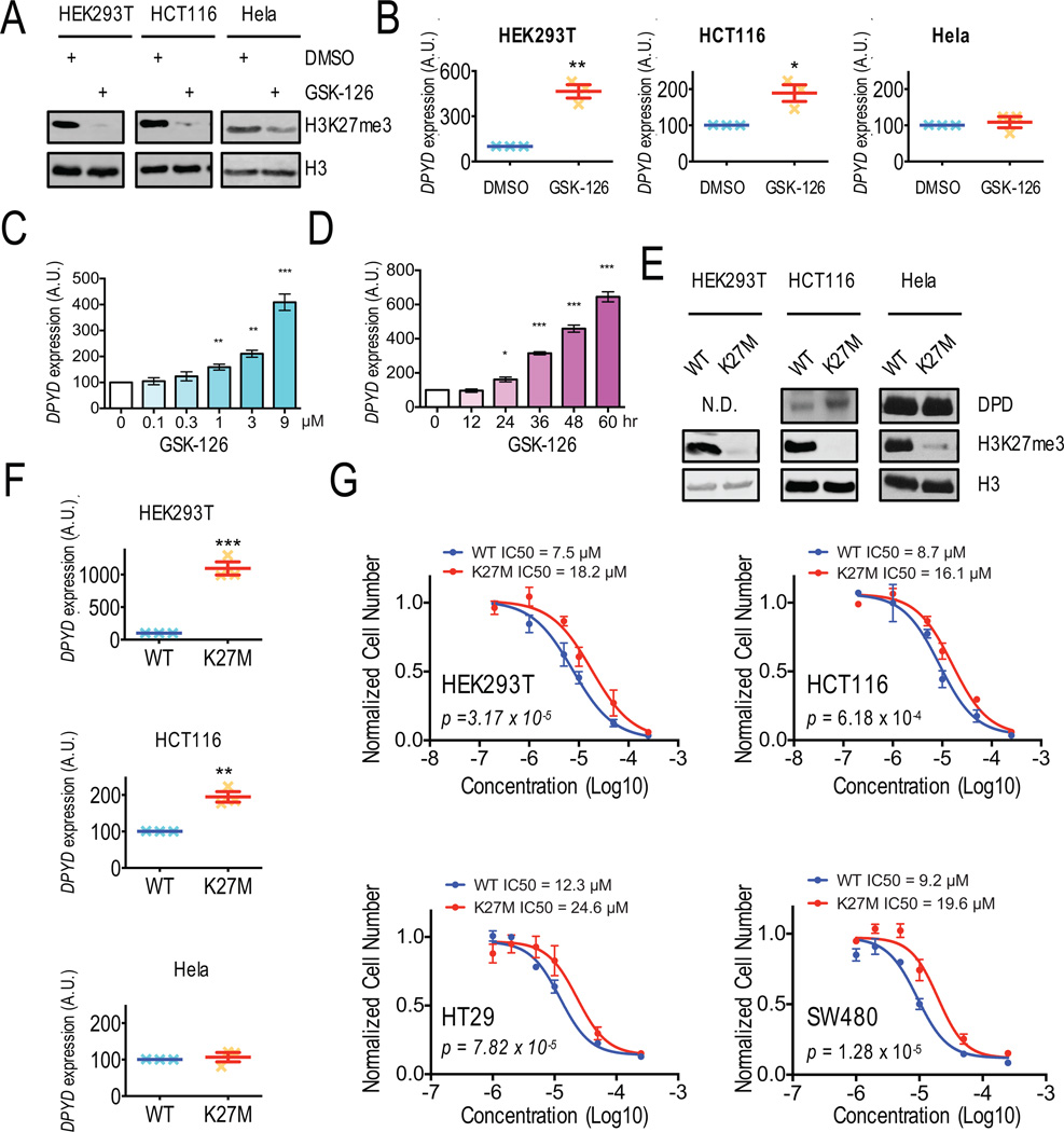
Figure 2. Inhibition of Ezh2 results in increased DPYD expression and desensitizes cells to 5-FU.
A. Western blotting was used to measure histone H3 and H3K27me3 levels in HEK293T, HCT116, and Hela cells treated with DMSO or 1 µM GSK-126. B. DPYD expression in cells from (A) was measured by q-RT-PCR. C. DPYD expression was measured in HEK293T cells treated with the indicated concentrations of GSK-126. D. DPYD expression in HEK293T cells was measured at indicated time points following treatment with 1 µM GSK-126. E. DPD, H3K27me3, and total H3 expression in HEK293T, HCT116 and Hela cells stably expressing wild-type H3 and H3K27M were measured by western blotting. F. DPYD expression was measured in cells from (E). G. IC50 concentrations for 5-FU were determined using HEK293T, HCT116, HT29 and SW480 cells stably expressing wild-type H3 (WT) or H3K27M (K27M). In all panels means +/− SD are represented by bars and whiskers. N.D., not detectable; * p<0.05; ** p<0.01; *** p<0.001; N.D. not detectable.

