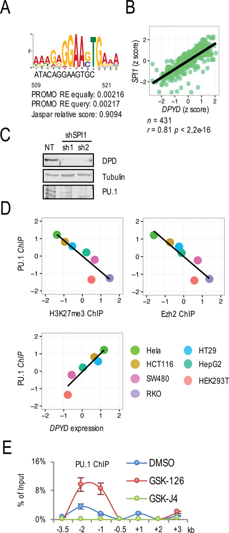
Figure 5. H3K27 trimethylation prevented PU.1 binding at the DPYD promoter.
A. The PU.1 binding site was identified on the DPYD promoter located 509–521 nucleotide upstream from the TSS using PROMO. B. Correlation between DPYD and SPI1 expression was determined in colon/rectum cancer TCGA data. C. DPD and PU.1 expression were measured by western blotting of HCT116 cells treated with two different shRNAs directed at PU.1 (sh1 and sh2) or non-target control shRNA (NT). D. Correlations between PU.1 and H3K27me3 enrichment at the DPYD promoter (top left), PU.1 and Ezh2 enrichment at the DPYD promoter (top right), and PU.1 and DPYD mRNA (bottom left) were determined for cell lines indicated (bottom right). Cumulative ChIP enrichment was calculated as in Figure 3E. E. Changes in PU.1 enrichment at the indicated positions (relative to DPYD TSS as indicated) were determined by ChIP-qPCR from HCT116 cells treated with 1 µM GSK-126, 1 µM GSK-J4, or DMSO control.

