
Figure 6. In vitro stimulation of the growth of FDC-P1 cells by different fractions of vesicles.
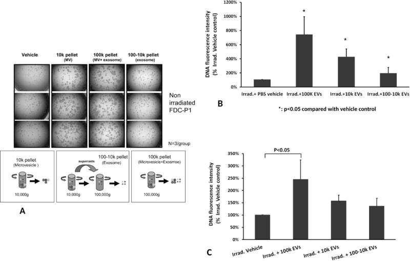
(A) The effects of different separative approaches on proliferation of non-irradiated FDC-P1 cells. Cell proliferation images were taken under microscopy with 2.5× objective (N=3/group). (B) Different fractions of WBM-EVs promoted radiated FDC-P1 cell proliferation. The proliferation of radiation damaged FDC-P1 cells was determined by using CyQUANT NF Cell Proliferation Assay, with values normalized to the levels of untreated cells. The cells were treated with three fractions of vesicles for 10 days (mean ± SD, n=3/group). *: P<0.05, compared to Irrad. + PBS vehicle control. (C) Effect of different human MSC-EVs fractions on irradiated FDC-P1 growth in vitro. FDC-P1 cells were co-cultured with 2×10⁹/ml of EVs after 500 cGy exposure. The proliferation of cells was determined by using CyQUANT NF Cell Proliferation Assay after 10 days of MSC-EVs treatment (mean ± SD, n=3/group).