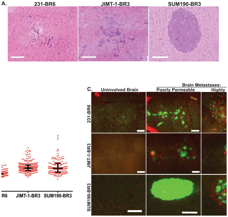
Figure 1. Heterogeneous permeability of three experimental brain metastasis model systems of breast cancer to 3kDaTexas Red dextran.
Tumor cells from three human brain-tropic cell lines were each injected into the left cardiac ventricle of immunocompromised mice; just before necropsy mice were injected with 3kDa Texas Red dextran. At necropsy, mice were perfused with saline such that the only Texas Red dextran remaining would be that permeable into tissue. A. Representative H&E stained sections of the triple negative 231-BR6, HER2+ JIMT-1-BR3 and HER2+ inflammatory breast cancer SUM190-BR3 brain lesions; scale bar=300 μm. B. Texas Red dextran diffusion of 143-256 metastases/model system was determined by quantitative immunofluorescence (see Methods and Supplemental Methods) and plotted as a ratio of metastasis to the BBB in uninvolved brain (set at 1.0). Lines indicate the median and the bars represent the upper and lower quartiles. Each dot represents a single metastasis (n= 5 mice). C. For quantitative immunofluorescence analysis of BBB and neuro-inflammatory response markers, highly and poorly permeable lesions within a single brain (tumor cells, green) were selected visually based on Texas Red diffusion. A representative photomicrograph of each is shown, along with uninvolved brain as a control; scale bar=200 μm (Red, Texas red dextran; Green, tumor cells). High permeability appeared as a diffuse cloud of red staining, or areas of diffusion around vessels.

