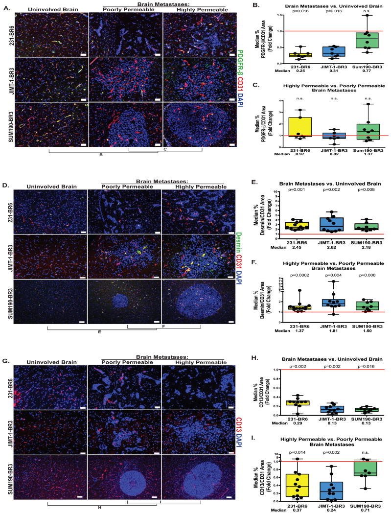
Figure 5. Desmin-positive pericytes identify highly permeable metastases.
For the three model systems described in Figure 1, metastases with distinctly high and low permeability to Texas Red dextran were identified within each animal brain, and adjoining tissue sections were stained for three markers of pericytes: PDGFR-β (A-C), Desmin (D-F) and CD13 (G-I). Quantitative immunofluorescence of each marker in uninvolved brain versus metastasis, and poorly versus highly permeable metastases was plotted as described in Figure 2. A-C. PDGFR–β+ pericytes (green) normalized to CD31 (A). PDGFR–β+ pericyte coverage was decreased in metastases as compared to uninvolved brain in the 231-BR6 and JIMT-1-BR3 model systems (B), but was unrelated to metastasis permeability in all three models (C); scale bar=50 μm. D-F. Expression of the desmin+ subpopulation of pericytes (green) normalized to CD31 (D) by the same methods (yellow = co-stain). The data show both a significant increase in desmin+ pericyte expression when brain metastases were compared to uninvolved brain (E) as well as increased expression in more permeable metastases as compared to less permeable metastases in the same brain (F); scale bar=50 μm (231-BR6 and JIMT-1-BR3) and 100 μm (SUM190-BR3). G-I. Expression of the CD13+ subpopulation of pericytes (G) normalized to CD31 (in an adjacent section) by the same methods. The data demonstrate both a decrease in CD13+ pericyte expression when brain metastases were compared to uninvolved brain (H), as well as a strong trend of further decreased expression in more permeable metastases as compared to less permeable metastases in the same brain in the 231-BR6 and JIMT-1-BR3 models (I); scale bar=50 μm (231-BR6 and JIMT-1-BR3) and 100 μm (SUM190-BR3).

