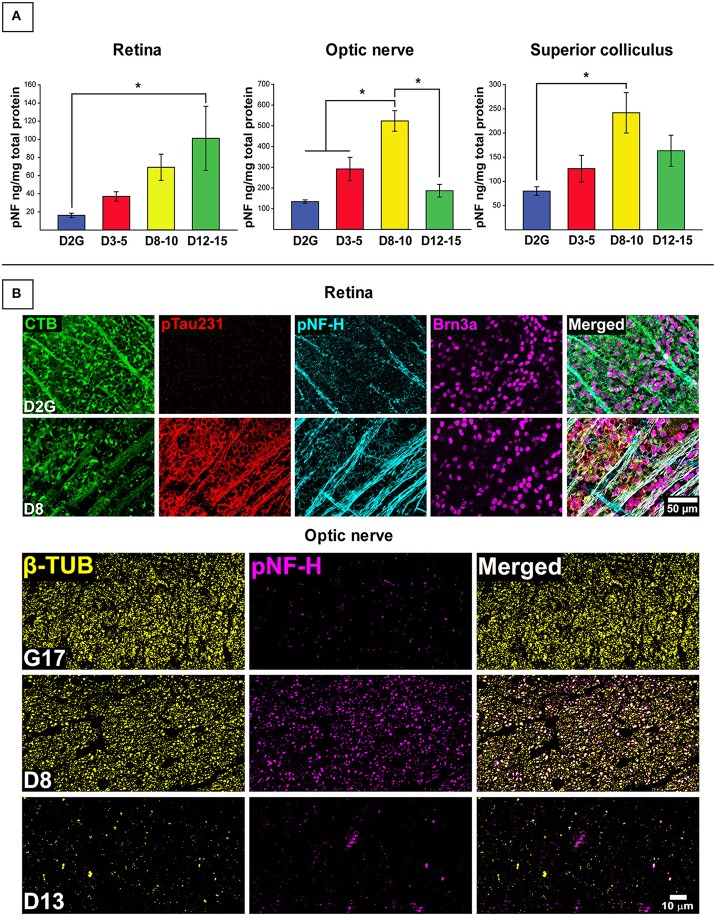
Figure 1.
Early elevations in pNF-H observed in distal RGC projection with effects of age and transport outcome. (A) Quantification of pNF-H by ELISA in retina, optic nerve (ON), and superior colliculus (SC). DBA/2J retinal pNF-H levels show trending increase with age peaking at 12–15 months at which point, is significantly elevated compared to the D2G controls. In the ON, pNF-H levels were highest at 8–10 months. in the DBA/2J mice but returned to levels similar to the D2G controls at 12–15 months. In the SC, pNF-H levels remained significantly elevated in the D8-10 group compared to controls. (B) (Top) Immunofluorescence of cytoskeletal markers, SMI-310 (specific for superphosphorylated, pNF-H) and the RGC marker Brn3a in the retina of an 8-mo. DBA/2J (D8) and D2G control. Micrographs show visibly increased pNF-H and somatic/axonal ptau-231 staining in the D8 retina. The D2G pNF-H labeling is typical of mature, non-pathological axons within the retina. (Bottom) Semithin (1 μm) cross-sections taken from 8-months. DBA/2J (D8), 13-months. DBA/2J (D13), and 17-months. D2G (G17) control ON immuno-stained for β-tubulin (β-TUB) and pNF-H. A significant increase in pNF-H is observed in the D8 ON compared to G17 control. In the D13, pNF-H in the ON is nearly absent corresponding to the prominent loss of axonal structure indicative by the reduction in β-TUB. Error bars depict SEM. Asterisk indicates significant statistical difference (p <0.05).

