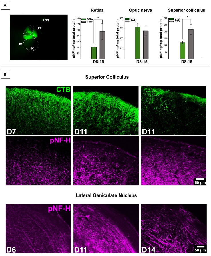
Figure 2.
Transport-dependent elevations in pNF-H observed in retina and SC of DBA/2J mice. (A) (Left) Photomicrograph from the dorsal surface of a whole-mount pathological SC in which the cortex has been removed and cholera toxin-B (CTB) labeling shows sectorial loss in the left (top) collicular lobe and complete loss in the right (bottom) lobe. Abbreviations: IC, inferior colliculus; PT, pre-tectum; LGN, lateral geniculate nucleus; SC, superior colliculus. The dotted lines demarcate areas absent of CTB indicating axonal transport deficit (CTB-). (Right) ELISA quantification of pNF-H in retina, optic nerve (ON), and SC. The transport status of retina and ON were determined by whether or not the SC was CTB+ (>90% CTB-label for partially transporting projections) or CTB- (<10% CTB-label for partially transporting projections). Levels of pNF-H were significantly elevated nearly 2-fold in both retina and SC of CTB- projections compared to CTB+ projections. Asterisks indicate significant statistical differences (p <0.05). Error bars depict SEM. (B) Top panel shows immunofluorescence of the SC, comparing 7-months. (D7), and 11-months. (D11) DBA/2J with and without CTB transport. Visibly increased pNF-H is shown in CTB- D11 SC. Bottom panel illustrates an age-dependent increase in pNF-H staining in the LGN (the visual thalamus) of 6, 11, and 14-month DBA/2J mice.

