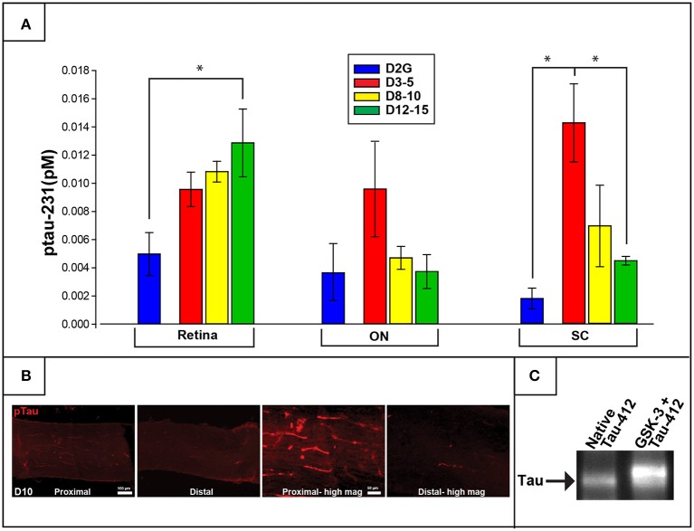
Figure 3.
Phosphorylated tau (ptau-231) is elevated early in the distal projection and is observed to translocate to the retina in pathologically-aged animals. (A) Multiplex quantification of ptau-231 in DBA/2J mice shows an age-dependent decrease of ptau in SC with an age-dependent increase in retinal ptau. In the SC, ptau-231 is significantly higher in the D3-5 group compared to D12-15 and D2G control group levels. In retina, however, D12-15 ptau-231 levels are significantly higher than D2G controls. Ptau levels in ON samples did not differ between groups. (B) Immunofluorescence for ptau-231 in 10-months. DBA/2J ON shows evidence of preferential accumulation of ptau-231 in the proximal portion compared to the distal portion of the nerve. (C) An example of co-immunoprecipitation data comparing native and hyperphosphorylated tau via reaction with GSK-3 suggests an increased affinity of phosphorylated tau for the retrograde motor component, dynactin-2. This is qualitatively illustrated by the increased optical density of Tau-5 (a marker for pan-tau) in the second lane representing hyperphosphorylated tau (GSK-3+Tau-412), compared to the first lane which contained only native tau. The tau band shown here corresponds to a molecular weight of approximately 43.5 kDa corresponding to the GSK-3 phosphorylation site, pS396/404. Asterisk indicates that groups are significantly different from bracketed comparison (p <0.05). Error bars depict SEM.

