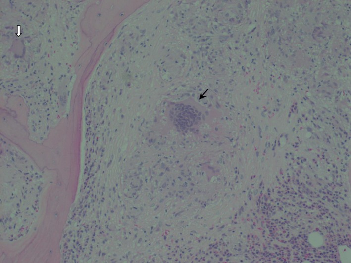
Figure 2.

The noncaseating granuloma in the center (black arrow) is surrounded by pale pink amorphous material with scattered mononuclear cells interspersed throughout with some giant cells. There is also a surrounding cuff composed of reactive cells such as eosinophils and neutrophils. A second granuloma is visible in the periphery containing a Langhans giant cell (white arrow). Hematoxylin and eosin stain (x20).
