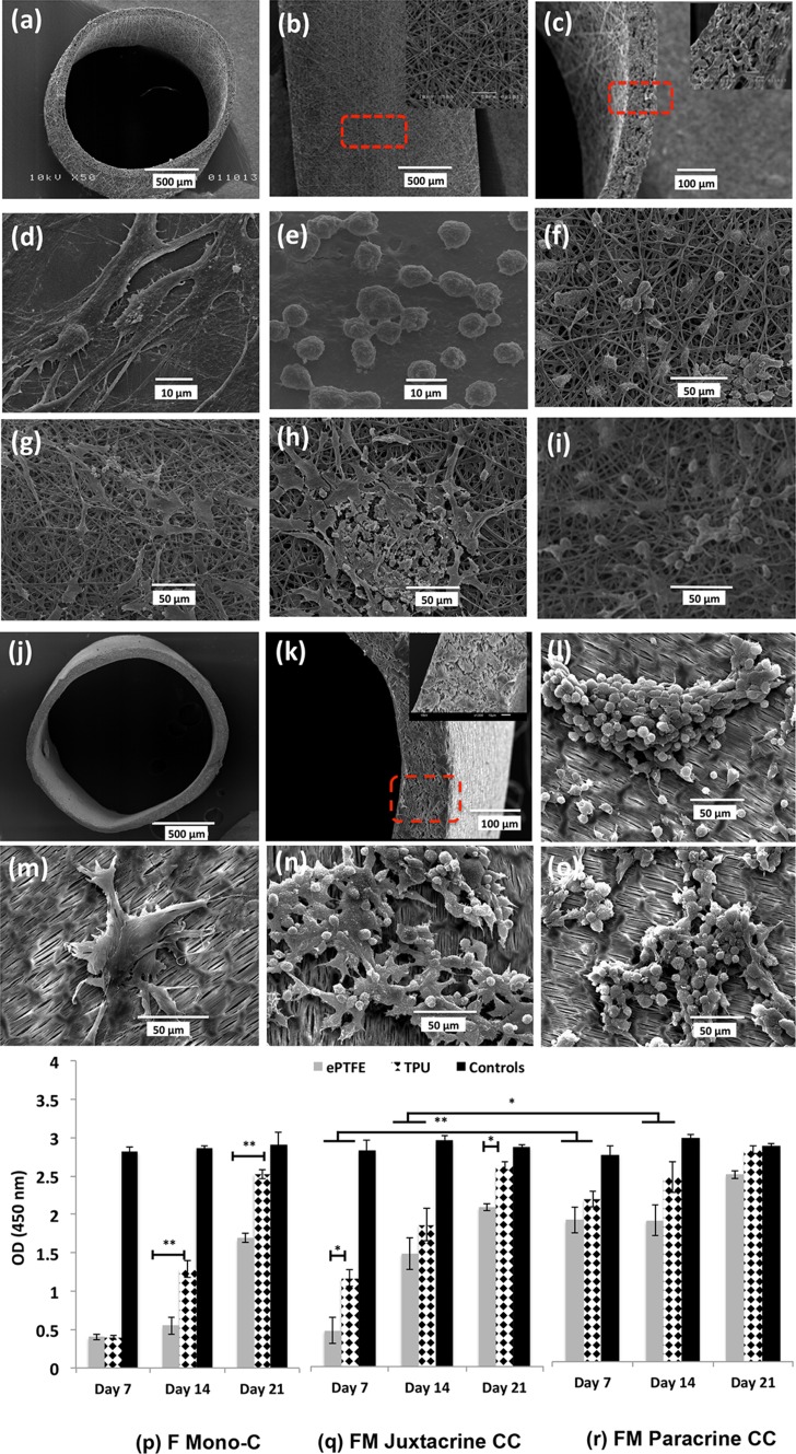
Figure 1.
Morphological and cell viability/proliferation studies. SEM micrographs of TPU/ePTFE grafts and morphology of the isolated primary fibroblasts and macrophages seeded on coverslips as control and on TPU/ePTFE grafts in mono-cultures and co-culture models, after 21 days. (a) TPU graft, (b) adventitial surface of the TPU graft and close up of the surface, (c) cross-section and close up of the wall structure, (d) primary fibroblasts (e) primary macrophages cultivated on plastic coverslips, (f) fibroblast mono-culture, (g) macrophage mono-culture, (h) fibroblast-macrophage juxtacrine co-culture and (i) macrophages in the fibroblast-macrophage paracrine co-culture after 21 days in TPU grafts. (j) ePTFE graft, (k) cross-section and close up of the wall structure, (l) macrophage mono-culture, (m) fibroblast mono-culture, (n) fibroblast-macrophage juxtacrine co-culture and (o) macrophages in the fibroblast-macrophage paracrine co-culture after 21 days, in ePTFE grafts. (p-r) Proliferation of fibroblast cells at different time points (7, 14 and 21 days), on TPU and ePTFE grafts in (p) fibroblast mono-culture, (q) fibroblast-macrophage juxtacrine co-culture and (r) fibroblasts seeded on transwell membrane in fibroblast-macrophage paracrine co-culture models. Data represent mean ± S.D. (n = 3 per time-point per group, technical replicates: 3). *p < 0.05, **p < 0.001.

