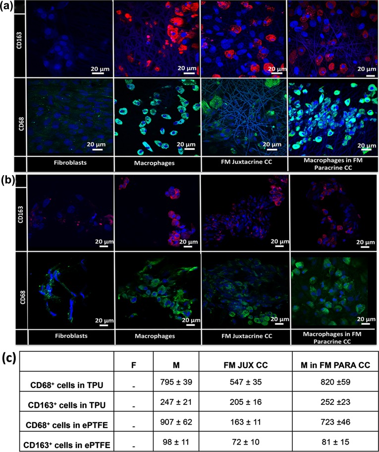
Figure 2.
Cell distribution studies and quantification of the positive cells via confocal microscopy. CD68 (green) and CD163 (red) immunofluorescence staining of macrophage and fibroblast mono-cultures and fibroblast-macrophage juxtacrine (FM Juxtacrine CC) and paracrine co-cultures (FM Paracrine CC) in (a) TPU and (b) ePTFE grafts after 21 days. The nuclei of cells were counterstained with DAPI (blue). (c) Quantification of the CD68 and CD163 positive cells on TPU and ePTFE grafts after 21 days in macrophage (M) and fibroblast (F) mono-cultures and fibroblast-macrophage juxtacrine (FM JUX CC) and paracrine co-cultures (FM PAR CC). Data are presented as mean ± standard deviation (n = 3 per group).

