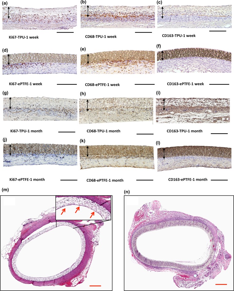
Figure 5.
Immunohistochemical staining for (a, d, g, j) Ki67, (b, e, h, k) CD68, (c, f, i, l) CD163 for TPU and ePTFE grafts after 1 week and 1 month implantation. Black arrows indicate graft walls, black scale bar: 200 µm. H&E stained of the cross-section of the (m) TPU and (n) ePTFE grafts after 1 month implantation, red arrows point out the luminal endothelial layer, red scale bar: 50 μm.

