
Figure 3.
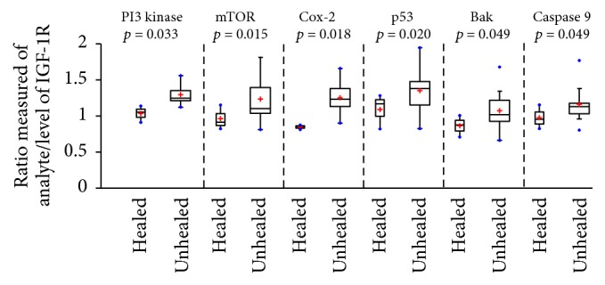
Boxplots showing significantly increased levels of downstream proteins compared to levels of IGF-1R in healed and unhealed DFU keratinocytes. Boxplots showing the calculated ratios for the level of each analyte/level of IGF-1R for healed (n = 3) and unhealed (n = 15) DFU subjects. These boxplots demonstrate that the unhealed subjects have significantly (p ≤ 0.05) elevated ratios of PI3 kinase, mTOR, Cox2, p53, Bak, and Caspase 9 compared to healed subjects. Not only is the center higher for unhealed subjects, but the quantitative independent two-samples t-test analysis suggests that the 2-population means differ beyond random variation.