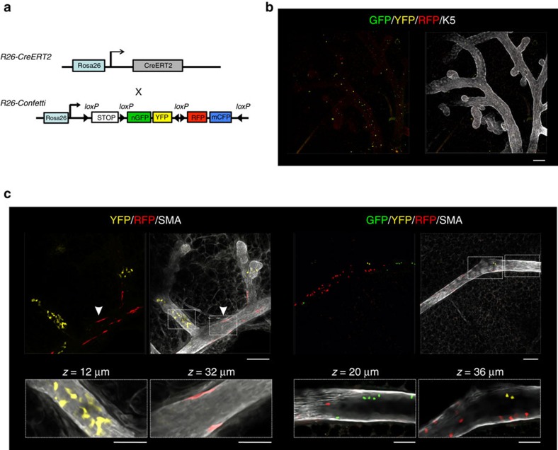
Figure 5. Clonal labelling patterns observed in R26-Confetti;R26-CreERT2 pubertal mice.
(a) Schematic representation of the R26-Confetti;R26-CreERT2 mouse model. (b) Initial labelling level observed 2 days after the administration of a single, low-dose of tamoxifen (1 mg intraperitoneal (i.p.)) to 4-week-old mice. Scale bar, 100 μm. (c) Labelling patterns observed in R26-Confetti;R26-CreERT2 pubertal mice confirm the results from the R26[CA]30 model. Labelling was induced by the administration of a single, low-dose of tamoxifen (1 mg i.p.) to 4-week-old mice and mammary glands were collected after a 3-week chase. Left panel shows a region containing YFP+ luminal cells and RFP+ basal cells (arrowhead) populating three branches, interspersed with unlabelled cells. Right panel shows a region containing GFP+, YFP+ and RFP+ luminal cells in a single branch. Scale bar, 100 μm (overview) and 50 μm (inset). Images are representative of three mice. Additional images are shown in Supplementary Fig. 11.

