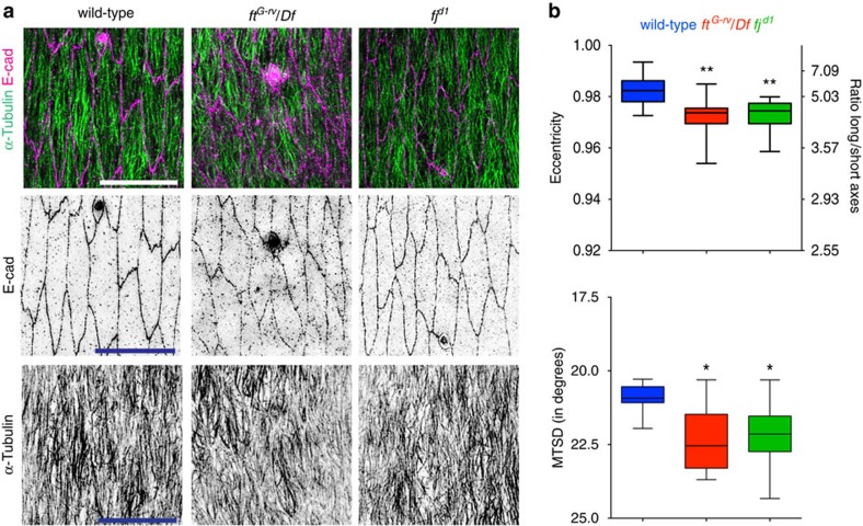
Figure 1. Loss of function of PCP pathway components Fat and Four-Jointed results in weak defects of cell shape and MT organization.
(a) Dorso-lateral regions of stage 15 embryonic epidermis, with apical cell outlines revealed with an E-cadherin (E-cad) antibody (magenta) and MTs with an α-Tubulin antibody (green): wild-type (left), fat mutant over fat deficiency (ftG-rv/Df; centre) and four-jointed mutant (fjd1; right). Scale bars, 10 μm (b) Quantification of cell eccentricity and cell's long/short axes ratio (top) and MT alignment (MTSD; bottom). Cells from 12 embryos from 3 (wild type, ftG-rv/Df) and 2 (fjd1) independent experiments were analysed. Statistical analysis of data combined with data from Supplementary Fig. 2a,b was performed with one-way ANOVA and Fisher's LSD post hoc analysis. *P<0.05, **P<0.01.

