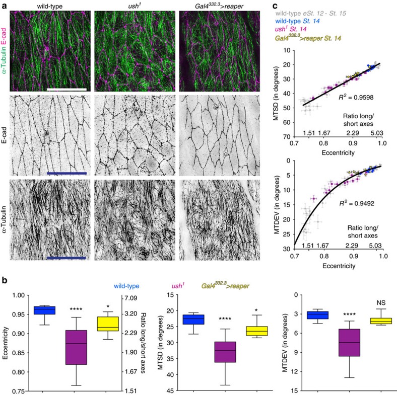
Figure 3. Changing cell shape alters sub-apical MT organization.
(a) Representative images of dorsal–lateral epidermis, visualized as described in Fig. 1, of stage 14 wild type (left) and embryos with germband retraction blocked by the ush1 mutant (centre) embryos, or inducing cell death in the amnioserosa using Gal4332.3>reaper (right). Scale bars, 10 μm. (b) Quantification of cell eccentricity and cell's long/short axes ratio (left), MTSD (centre) and MTDEV (right) shows germband retraction is required for cell elongation and MT organization. Cells from 12 embryos (wild-type, ush1 mutant) and 10 embryos (Gal4332.3>reaper) from two independent experiments were analysed. Statistical analysis was performed with one-way ANOVA and Fisher's LSD post hoc analysis. *P<0.05, ****P<0.0001, NS, not significantly different. (c) Correlation between either MTSD (top) or MTDEV (bottom), and cell eccentricity is retained when cell elongation is blocked. Each dot represents the cells from a single embryo (mean±s.e.m.).

