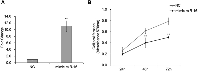
Figure 4. miR-16 suppressed embryonic fibroblast proliferation.
(A) Overexpression of miR-16 in DF-1 chicken embryo fibroblast cells using mimic miRNA. 20 nM mimics miR-16 and negative random RNA were transfected into cell lines, separately. After 36 h post transfection, the cells were harvested for detecting gene expression (n = 3). U6 was used as the reference gene. Fold change values were calculated using the 2−∆∆Ct method. (B) The affection of miR-16 on DF-1 chicken embryo fibroblast cell growth. 20 nM mimic miR-16 and negative random RNA were transfected in DF-1 chicken embryo fibroblast cells, respectively (n = 6). After different periods of culturing (24 h, 48 h and 72 h), the ability of cell proliferation was assayed with MTT assay. The proliferation curves were calculated values of optical density at 570 nm. The curves show that miR-16 significantly inhibited DF-1 chicken embryo fibroblast cell proliferation. The data are presented as the means ± SD. *p < 0.05 and **p < 0.01 were estimated by Student’s t-test.

