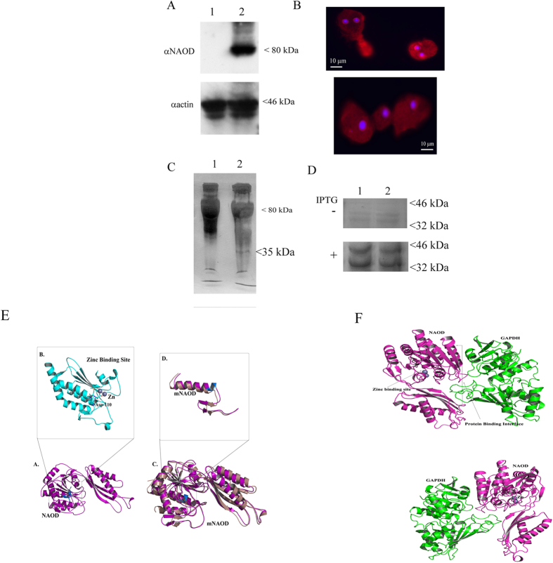
Figure 7. NAOD and GAPDH interact.
(A) Western blot analysis of whole protein extract prepared from HaloTag control trophozoites (lane 1) and from HaloTag-NAOD trophozoites (lane 2). The proteins were separated on 10% SDS-PAGE gels and analyzed with an NAOD antibody (αNAOD) or an actin antibody (αactin). The cropped image displays a representative result from three independent experiments. Full length blot is presented in Supporting Information S4. (B) Immunofluorescence confocal microscopy of HaloTag-NAOD trophozoites. HaloTag-NAOD (red) was detected using HaloTag TMR Ligand (1 μM). The nuclei (blue) were stained by DAPI. No fluorescence was observed when Halo-tagged trophozoites were incubated with HaloTag TMR Ligand. (C) HaloCHIP of NAOD from HaloTag-NAOD trophozoites exposed of not to GSNO (350 μM for 30 min). A 35 kDa protein is specifically pull-down from the whole protein extract of HaloTag-NAOD trophozoites. (D) NAOD (or mutNAOD) and GAPDH are co-expressed from the pETDuet-1 expression vector in E. coli. In this vector, NAOD (lane 1) or mNAOD (lane 2) was expressed as a His-tagged protein and GAPDH was untagged. His-tagged NAOD or mNAOD was pull-downed from non-IPTG-induced E. coli or IPTG-induced E. coli lysates with Ni-NTA resin. The pull-downed proteins were resolved on an SDS-PAGE gel and the gels were Coomassie stained. (E) Model of NAOD (purple color). (A) The structure shows the binding of Zinc at the active site along with interacting residues. (B) Zoomed view of the zinc binding site (cyan color) along with the ASP-110 in stick model. (C) Superimposition of mNAOD (violet color) with full length NAOD. (D) Zoomed view of the zinc binding site in mNAOD structure compared to the full length model. (F) Model of NAOD-GAPDH interaction. (upper panel). Final complex (best pose) obtained from the docking simulation of NAOD-GAPDH interaction. The figure shows the binding site of Zinc and the protein-protein interaction site between NAOD and GAPDH. (lower panel). 180 degree rotated view of the interaction between NAOD and GAPDH.

