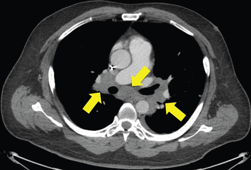
Figure 2.

CT of hilar lymphadenopathy, which was initially believed to be tumour progression. After three doses of immunotherapy, restaging CT scans showed interval enlargement in the known tumour mass in the bed of kidney resection and new hilar lymphadenopathy. Immunotherapy was discontinued due to disease progression on CT scans, and he began chemotherapy with gemcitabine and cyclophosphamide. However, a hilar lymph node biopsy via bronchoscopy showed no evidence of cancer but was consistent with sarcoid.
