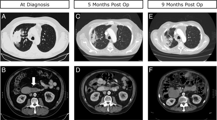
Figure 1.
(A) Preoperative CT scan of the chest demonstrating a large primary lesion in the patient's right upper lobe. (B) Preoperative CT scan of the abdomen demonstrating a hypodense, fully obstructive metastatic lesion in D3, identified by the white arrow. (C) Five-month postoperative CT scan of the chest demonstrating reduction in the size of the primary lung tumour. (D) Five-month postoperative CT scan of the abdomen demonstrating limited interval change in the metastatic lesion in D3. (E) Nine-month postoperative CT scan of the chest demonstrating moderate progression of the primary lung lesion. (F) Nine-month postoperative CT scan of the abdomen demonstrating distension of D2.

