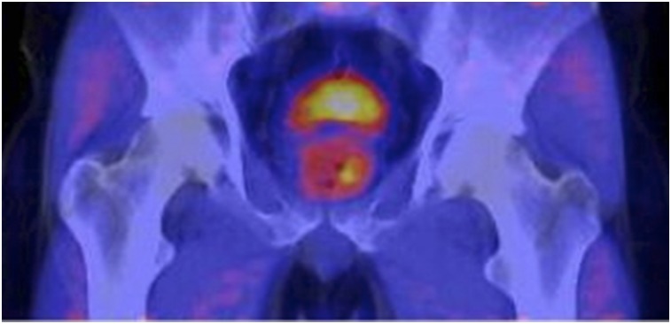
FIGURE 4.
61-y-old man who had elevated serum PSA level (10.5 ng/mL) and had undergone standard transrectal ultrasound biopsy, with negative results. Axial PET/CT with 18F-FMAU demonstrated focally increased tracer uptake in left base of prostate gland. PET/multiparametric MRI–directed biopsy revealed atypical small acinar proliferation suggestive of early malignancy.

