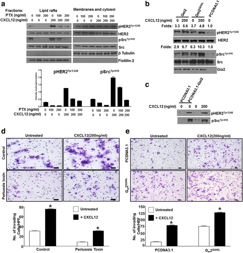
Fig. 2.

CXCL12 activation of HER2 and Src is mediated by Gαi GTP proteins in lipid raft membrane microdomains and this activation induces cell invasion. a C4-2B cells were cultured in the presence or absence of increasing concentrations of pertussis toxin (PTX) for 3 h, followed by treatment with CXCL12 for 15 min. Cell lysate was collected, and protein expression of phosphorylated and total HER2 and Src was determined by Western blot analysis. Flotillin was utilized as a loading control for the lipid raft fraction; β-Tubulin was utilized as a control for the membranes and cytosol fractions. Bottom panel shows the quantitation of phosphorylated HER2 and Src. b C4-2B cells were transfected with wild type Gαi (WT-Gαi2), constitutively active Gαi (Q205L), or plasmid control (pcDNA3.1). Cells were then cultured in the presence or absence of CXCL12 for 15 min, and cell lysate was collected. Protein expression of phosphorylated and total HER2 and Src as well as Gαi2 were determined by Western blot analysis. c C4-2B cells were transfected with plasmid control or constitutively active Gαi (Q205L) and cultured in the presence or absence of CXCL12. Lipid raft fractions were collected and immunoblotted with phospho HER2 and Src antibodies. d C4-2B cells were either treated or not, with PTX (200 ng/ml) and a chemoinvasion assay was performed in the presence or absence of 200 ng/ml CXCL12. e C4-2B cells were transfected with pcDNA3.1 or pcDNA3.1 Gαi2Q205L plasmids and a chemoinvasion assay was performed in the presence or absence of 200 ng/ml CXCL12. Bottom panels show the quantitation of invaded cells. Experiment was performed in triplicates; a representative field of invading cells are shown (d and e). Number of invading cells was quantitated in three independent experiments and statistical analyses were performed using ANOVA; significance was calculated using Tukey Posttest analysis using GraphPad Prism software. P value <0.05 is shown as a *
