
Fig. 2.
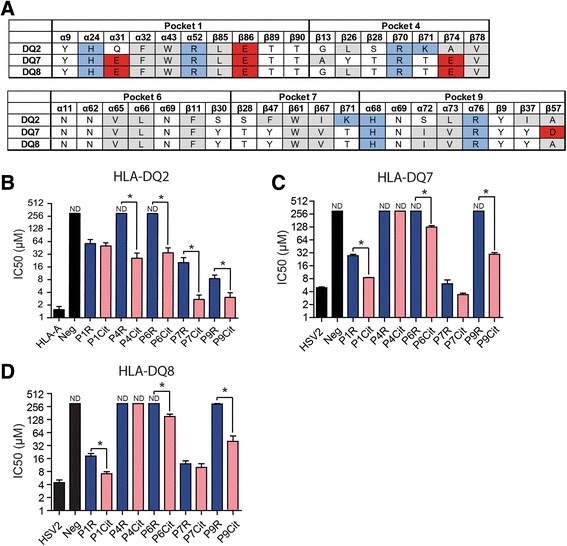
Accommodation of citrulline and arginine residues by HLA-DQ molecules. a Schematic representation of the differences in peptide-binding pockets between HLA-DQ2, HLA-DQ7 and HLA-DQ8. Amino acid (AA) residues are color coded according to their properties (white = hydrophilic, gray = hydrophobic, red = acidic, blue = basic). b Competitive binding of a biotin-labeled alpha-gliadin peptide with an unlabeled alpha-gliadin peptide or alpha-gliadin variants with citrulline or arginine residues in p1, p4, p6, p7, and p9 to HLA-DQ2. c–d Competitive binding of a biotin-labeled VP16 peptide with an unlabeled VP16 peptide or VP16 variants with citrulline or arginine residues in p1, p4, p6, p7, and p9 to HLA-DQ7 (c) and HLA-DQ8 (d). Graphs depict the IC50 values (μM). ND non-detectable binding. Binding experiments were performed at least three times and plots show pooled experiments. The error bars show the standard error of the mean. *Indicates a p value of <0.05