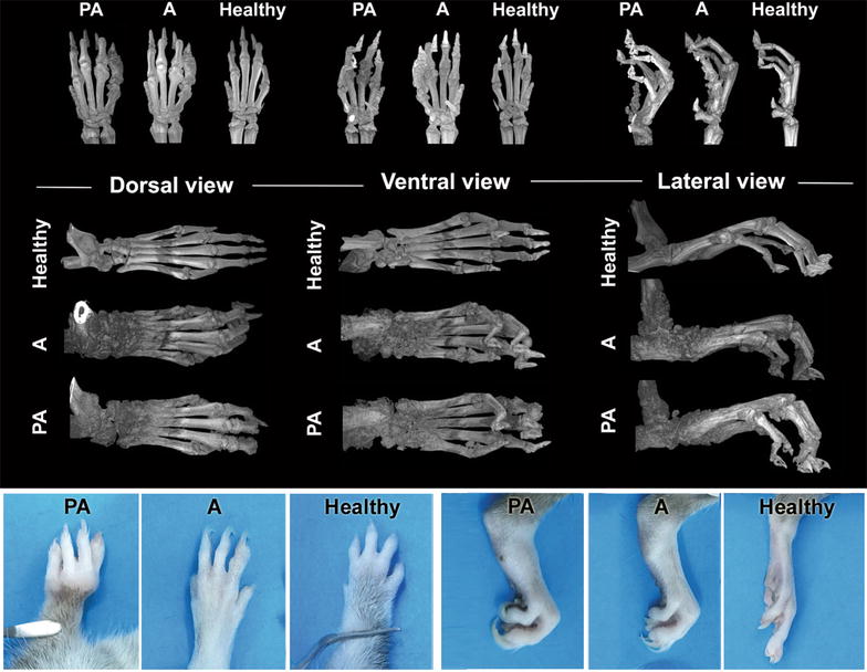
Fig. 3.

Micro-CT images and visual appearance of front and hind paws for assessment of arthritic changes. Micro-CT images of bone and tissue changes in front and hind paws of animals affected with experimental periodontitis and induced arthritis (PA), arthritis without periodontitis (A) and corresponding images of DA rats without induced arthritis or periodontitis (Healthy) at the time-point of 15 weeks; and visual appearance of front and hind paws at endpoint
