Fig. 1.

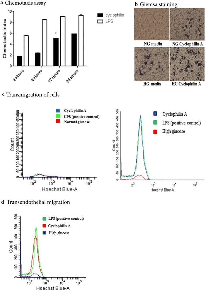
a Chemotactic response was analyzed using transwell assays as described in the method section. THP-1 cells were treated with or without cyclophilin A (100 ng/mL) for 4, 6, 12, 24 h. LPS (10 µg/mL) was taken as positive control. b Monocytes were cultured on the upper chamber of transwells in normal glucose (NG) and high glucose (HG) in the presence and absence of cyclophilin A (lower chamber) for 24 h. The adhered monocytes were stained using Giemsa stain. HG indicates RPMI 1640 culture media primed with high glucose (20 mM/L). (c) Flow cytometry analysis of cells after transmigration assays. Transwell experiments were performed using 5.0 micron pore membrane. To test the migration rate, THP 1 cells were seeded on top of the transwell and 100 ng/mL cyclophilin A was added to the bottom of chambers along with NG (left panel)/HG (right panel) media. Migration rate of THP cells cultured in HG treated with cyclophilin A were similar to cells treated with LPS alone indicating a strong chemokinetic activity by cyclophilin A. d Transendothelial migration of THP 1 cells across monolayer of endothelial cells (EaHy). THP-1 cells were seeded on top of the transwell and 100 ng/mL cyclophilin A was added to the bottom of the chamber. After 24 h of incubation transmigrated cells were counted by flow cytometry using Hoechst (10 µg/mL) as nuclear stain. Transendothelial migration of cyclophilin A treated cells were almost equal to that of positive control, LPS. Data are presented as mean ± SD (n = 3). Chemotactic response was analyzed using two-way ANOVA. p < 0.005 was considered significant
