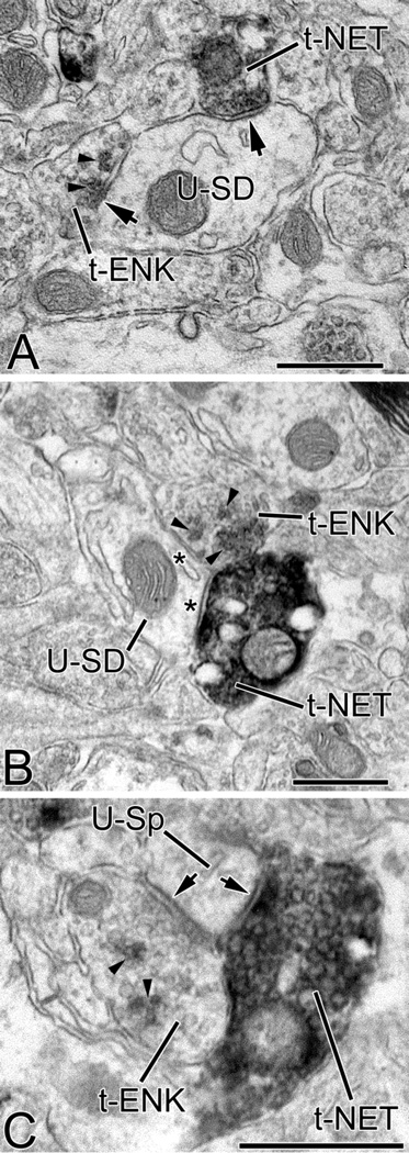
Fig. 2.
(A) High power photomicrograph of ENK-ir in the BLa in a section stained with nickel-intensified DAB. This field contains two ENK+ nonpyramidal neurons that are in the plane of focus, and numerous small ENK+ puncta presumed to be axon terminals; an axon with large varicosities (1 µm in diameter) is indicated with an arrow. (B) High power photomicrograph of ENK-ir in the lateral nucleus in a section stained with nickel-intensified DAB. This field contains numerous ENK+ puncta that average about 1 µm in diameter and appear to be axon terminals. (C) Four small ENK+ nonpyramidal neurons in the BLa in a section stained with non-intensified DAB. Small ENK+ puncta can be seen in the neuropil. (D) NET+ axons in the BLa. All scale bars = 25µm.

