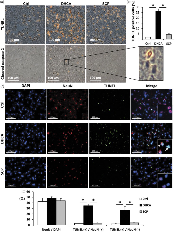
Figure 3.
Apoptosis cells and neurons identified by TUNEL staining in representative cerebral cortex sections. ((a), (b)) Detection of apoptosis and quantitation of apoptotic cells (brown/orange stains) from TUNEL staining. TUNEL assay showed no apoptosis in control pigs (Ctrl). Pigs undergoing DHCA had visibly more apoptosis compared with the SCP group, which also exhibited few apoptosis cells. Cleaved caspase-3 immunostaining (brown/orange stains) also showed same results as TUNEL staining. ((c), (d)) Double immunofluorescence staining for TUNEL (green) and the neuronal marker NeuN (red). Nuclei were counterstained with DAPI (blue). TUNEL-positive cells in DHCA group were detected in both neurons and other cells at the similar rate. TUNEL- and NeuN-double positive cells (arrowhead). TUNEL-positive and NeuN-negative cells (arrow). TUNEL, the terminal deoxynucleotidyl transferase-mediated dUTP nick end labeling. Scale bar = 100 µm. *p < 0.001.

