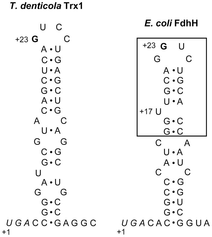
Figure 2. Structures of SECIS elements of T. denticola Trx1 and E. coli formate dehydrogenase H.
The SECIS of T. denticola Trx1 contains a conserved G nucleotide (bold) in the apical loop, but lacks a bulged U, which is present in the minimal step-loop structure (boxed) in E. coli formate dehydrogenase H (FdhH) SECIS. Sec UGA codons are shown in italics and numbering of nucleotides is stated at these Sec codons. The SECIS structure of T. denticola Trx1 was predicted using RNAfold [24].

