Figure 2.

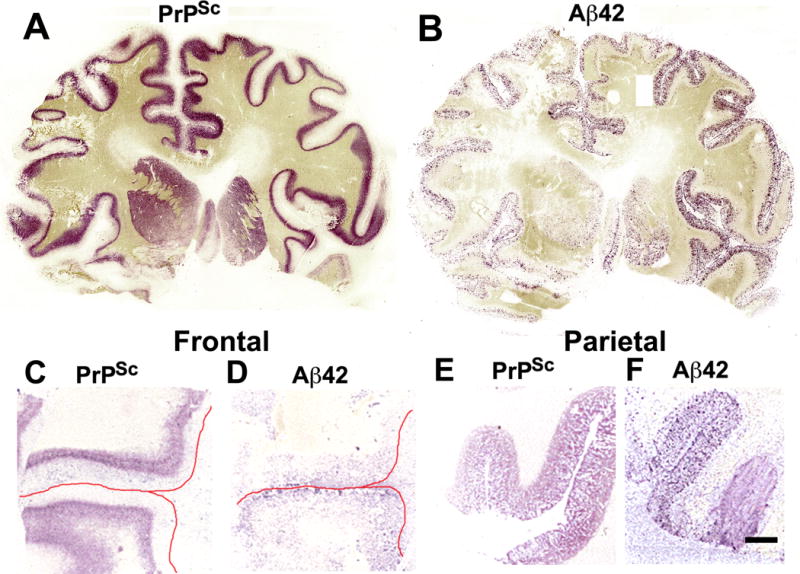
Scrapie-associated prion protein (PrPSc) and β-amyloid (Aβ42) histoblot analyses of the cerebrum in sporadic Creutzfeldt-Jakob disease (sCJD). (A, B) Full coronal sections from a sCJD case with incipient Alzheimer disease (AD) (case # 8, Table 1), not strictly fulfilling the Consortium to Establish a Registry for Alzheimer’s Disease (CERAD) criteria for AD. The 3F4 immunostaining shows PrPSc located mostly in cortical layers 5–6 (A). The 4G8 immunostaining indicates that Aβ42 plaques are located mostly in layers 1–4 of the cerebral cortex. A few Aβ42 plaques are also located in the caudate nucleus and putamen, particularly on the right side (B). (C, D) Sections of the lateral frontal cortex in another sCJD case with incipient AD (case #13, Table 1). PrPSc also localizes to cortical layers 5–6 (C). A few Aβ42-positive plaques are located in layer 1. The red lines mark the location of the pial surface (D). (E, F) Sections of the parietal cortex of the same case shown in (C) and (D). PrPSc (E) and Aβ42 plaques (F) are located in layers 1–6. Scale bar in (F) represents 4 mm and also applies to (C–E).
