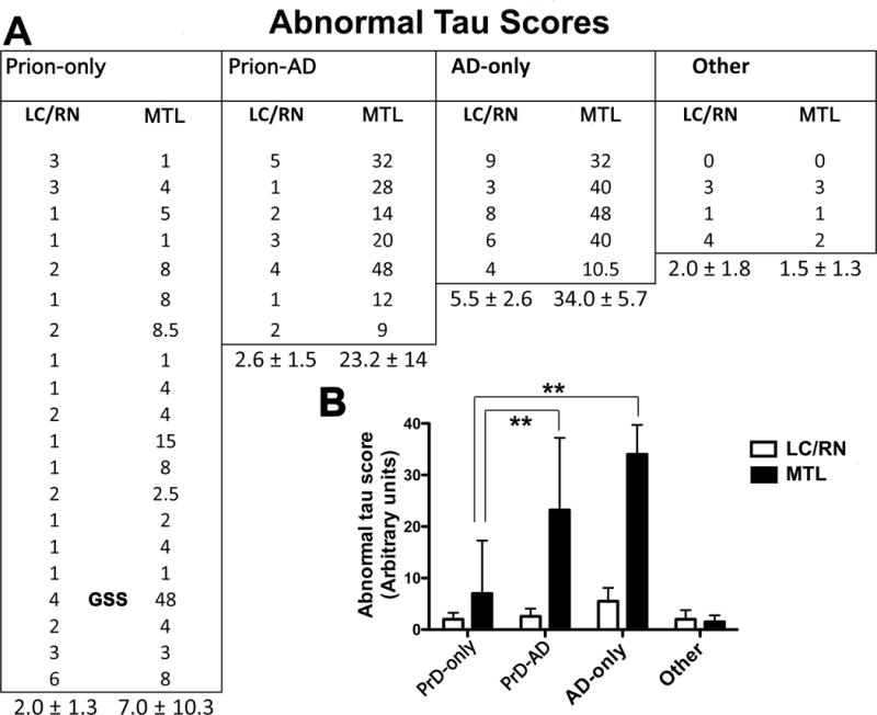
Figure 7.

Quantification of hyperphosphorylated tau protein (Hτ) immunostaining in the locus coeruleus and raphe nuclei (LC/RN) and the medial temporal lobe (MTL) in autopsy subjects, 4 of which were shown in Figure 6. (A) The estimated amount of Hτ-containing neuropil threads, nerve cell bodies, and dystrophic neurites were measured by densitometry in 20 Prion disease-only (PrD-only), 7 Prion-Alzheimer disease (Prion-AD), 5 AD-only, and 4 “Other” (3 TDP-43 encephalopathies and 1 dementia lacking distinctive abnormalities) cases. Means and standard deviations are given for the values in each category. (B) Abnormal tau scores are shown as a function of each disease group (mean ± SD). When values in the MTL were compared, abnormal tau was significantly higher in the AD-only and Prion-AD groups vs. the Prion-only group (19 CJD and 1 GSS marked in the figure) (**p < 0.001, t-test). GSS, Gerstmann-Sträussler-Scheinker disease.
