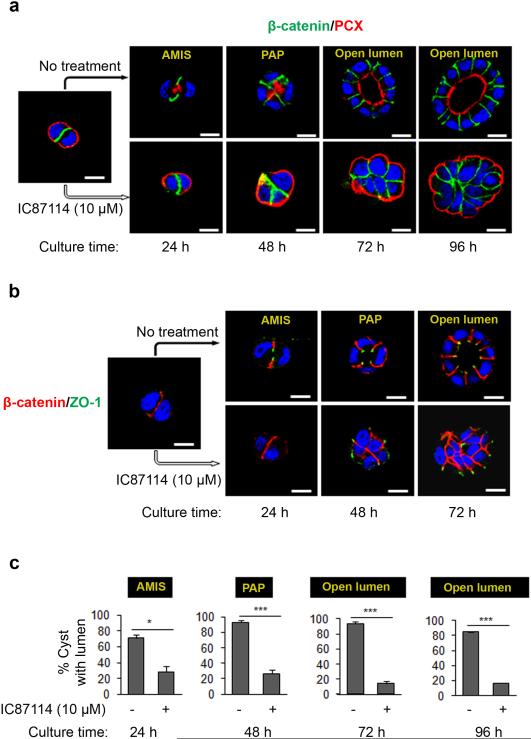
Figure 2. p110δ activity is required for lumen initiation in MDCK cysts.
(a) Time course analysis of polarity and lumen formation in MDCK cells, treated or not with 10 μM of IC87114 and plated on Matrigel to form cysts, fixed and stained after 24, 48, 72 and 96 h of culture, corresponding to different stages of lumen formation: AMIS = Apical Membrane Initiation Site (AMIS). PAP = Pre-Apical Patch and open lumen. Cells were stained for β-catenin (green) and PCX (red) and a single confocal section through the middle of cyst is shown. The scale bar represents 10 μm. (b) Cells were grown as in (a) and analyzed after 24, 48, and 72 h of culture and stained for β-catenin (red) and ZO-1 (green) and a single confocal section through the middle of cyst is shown. The scale bar represents 10 μm. (c) Percentage of MDCK cysts with AMIS, PAP or open lumen from 3 independent experiments were presented in histograms. values are expressed as mean ± s.e.m . Asterisk indicates student t test P<0.05, triple asterisks indicate P< 0.0001.

