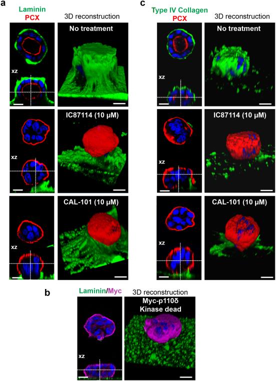
Figure 5. Laminin and type IV collagen assembly is dependent on p110δ activity.
(a) MDCK cells treated or not with 10 μM of p110δ inhibitors (IC87114 or CAL-101) were grown on matrigel for 4 days to form cysts and then fixed and stained with laminin (green), PCX (red) and Hoechst (blue) as indicated on the pictures. A confocal section through the middle of the cyst is shown and the XZ section is presented below each panel. The scale bar represents 10 μm. 3D reconstruction from all XZ sections is presented in the right column. (b) MDCK cells transfected or not with 1 μg of Myc-p110δ KD were grown on matrigel for 4 days to form cysts and then fixed and stained with laminin (green), Myc (purple) and Hoechst (blue) as indicated on the pictures. A confocal section through the middle of cyst is shown and XZ section is presented below each panel. The scale bar represents 10 μm. 3D reconstruction from all XZ sections is presented in the right column. (c) cells treated as in (a) stained with type IV collagen (green), PCX (red) and Hoechst (blue) as indicated on the pictures. A confocal section through the middle of the cyst is shown and the XZ section is presented below each panel. The scale bar represents 10 μm. 3D reconstruction from all XZ sections is presented in the right column.

