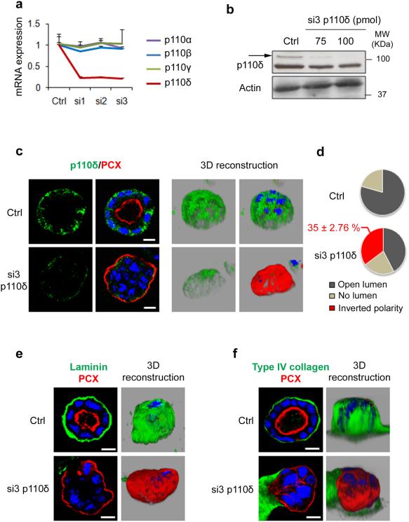
Figure 7. p110δ knock down alters laminin and type IV collagen assembly and leads to inverted polarized cyst formation.
(a) MDCK cells were transfected with 100 pmol of three different p110δ-specific siRNA (si1, si2 and si3) for 48 h. Total RNA was analyzed by qRT-PCR for p110δ, p110α, p110β and p110γ and normalized to GAPDH (see Methods). Control (Ctrl) corresponds to cells treated with transfection reagents only. values are expressed as mean ± s.e.m of four triplicate independent experiments. (b) Immunoblot analysis of p110δ using p110δ-specific antibody (sc7176), in MDCK cells transfected or not with 75 and 100 pmol of p110δ siRNA. Actin was used as a loading control. (c) MDCK transfected as in (a) with si3 or ctrl cells were grown in Matrigel for 96 h to form cysts and then stained for p110δ (green) using p110δ-specific antibody (ab32401), PCX (red) and Hoechst (blue). Single confocal section through the middle of the cyst is shown. The scale bar represents 10 μm. 3D reconstruction from all XZ sections is presented in the right column in grey background. (d) Quantification of open lumen, no lumen and inverted polarized cysts from 3 independent experiments was presented in pie. The percentage ± s.e.m. of inverted polarized cyst is indicated above the pie. (e) MDCK cysts in (c) were stained for laminin (green), PCX (red) and Hoechst (blue). A confocal section through the middle of cyst is shown. The scale bar represents 10 μm. 3D reconstruction from all XZ sections is presented in the right column in grey background. (f) MDCK cells in (c) were stained for type IV collagen (green), PCX (red) and Hoechst (blue). A confocal section through the middle of cyst is shown. The scale bar represents 10 μm. 3D reconstruction from all XZ sections is presented in the right column in grey background.

