
Figure 3. Fog2 is expressed by postmitotic corticothalamic projection neurons, but is excluded from corticospinal and callosal projection neurons.
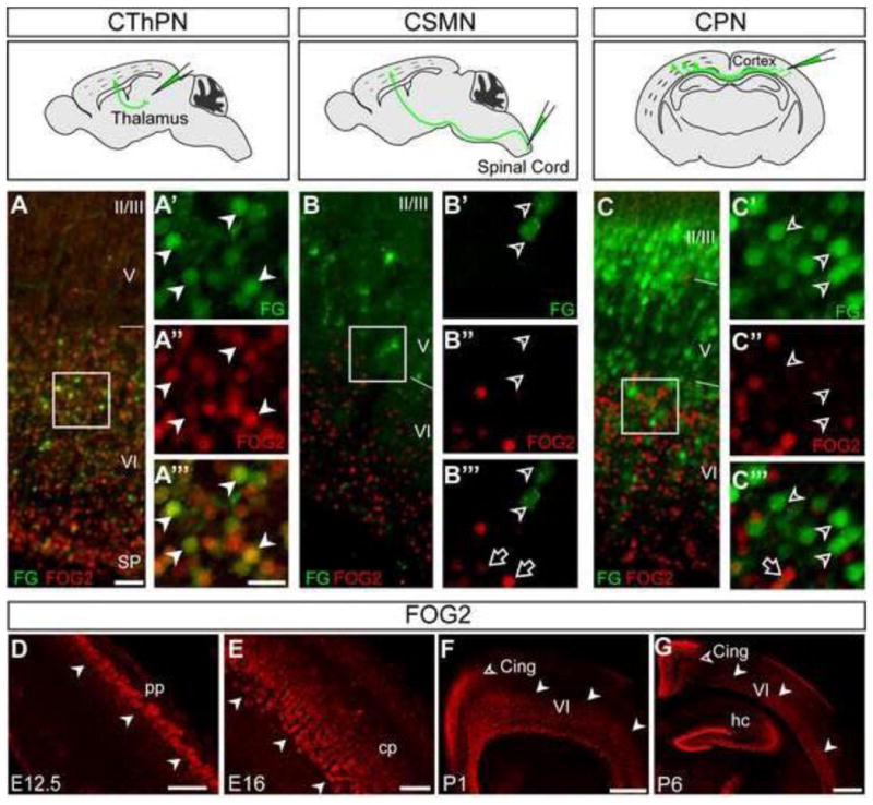
(A–C‴) FOG2 ICC (red) on coronal sections from brains injected with FluoroGold (FG) in thalamus, spinal cord, or contralateral cortex to retrogradely label CThPN, CSMN, or CPN, respectively (green). Schematic views of FG injection sites are at the top of each panel group. (A-A‴) FOG2 co-localizes with FG in retrograde labeled CThPN (white arrowheads in A′-A‴, area boxed in A). FG-labeled CSMN (B-B‴), or CPN (C-C‴) do not express FOG2. (D–G) Time-course of FOG2 expression revealed by ICC. FOG2 is first expressed at E12.5 in the preplate (white arrowheads) (D). FOG2 is strongly expressed in deep cortical plate during embryonic development (E), and in layer VI during the first postnatal week (white arrowheads) (F, G). Expression in the cingulate cortex is detected postnatally (open arrowheads F–G) Scale bars, 50 μm (A, B, C), 20 μm (A′-A‴, B′-B‴, C′-C‴), 50 μm (D–E), 200 μm (F), 500 μm (G).