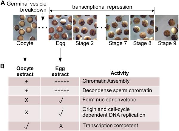
Figure 1. Xenopus laevis early embryology and extract functionality.
A. Stereomicrographs of Xenopus laevis stage IV oocytes, eggs (stage 1), and Stage 2 through Stage 10 embryos as indicated. Timing of germinal vesicle breakdown (GVBD) and the onset of transcriptional repression is indicated.
B. Cell-free oocyte extract is prepared from a pool of dissected oocytes (stages II-VI, primarily the later stages as the early stage oocytes are lost), while cell-free egg extract is prepared from laid eggs. The table shows various activities present in the extracts (+ = modest activity; +++++ = high activity; X = no activity; check = activity).

