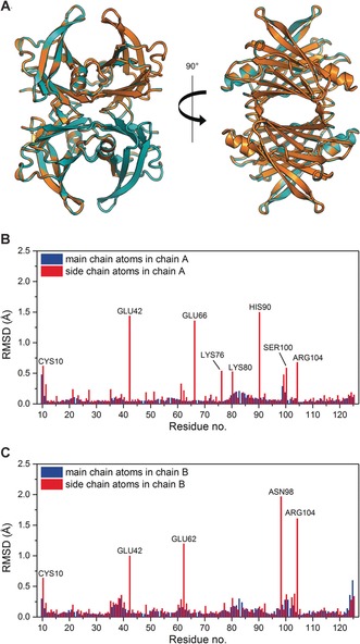
Figure 1.

A) Superposition of the X‐ray crystal structures of HTTR (orange color) and DTTR (teal color) show close similarity of the two variants, RMSD=0.11 Å over 908 main chain atoms. Image was generated and rendered with Pymol. Panels (B) and (C) show the calculated RMSD of the main chain atoms (blue) and side chain atoms (red) between HTTR and DTTR for residues in chain A and chain B, respectively. The residues Cys10, Glu42, Glu66, Lys76, Lys80, His90, Ser100, and Arg104 in chain A, and the residues Cys10, Glu42, Glu62, Asn98, and Arg104 in chain B are disordered and have poor density in the crystal structure analysis.
