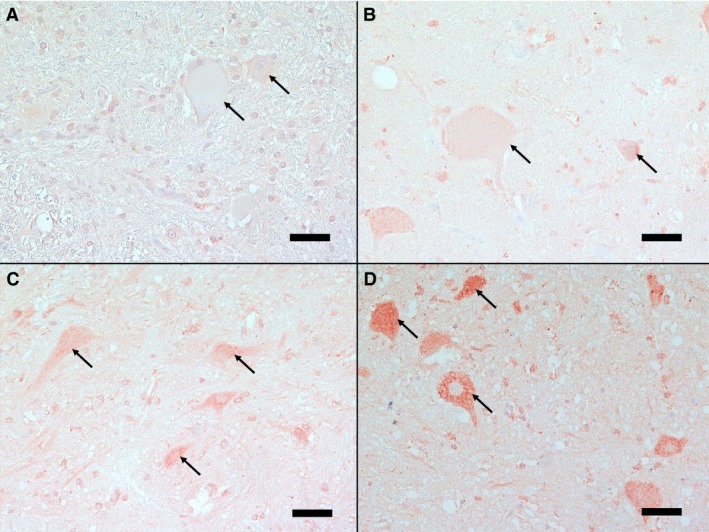
Figure 1.

Spinal cord cross‐sections showing different intensities of immunohistochemical staining for endothelin‐1 in neurons. Standard sections of different staining intensities of neurons (arrows) are presented: A‐ Grade 0: no positive staining, B‐ Grade 1: mild positive staining, C‐ Grade 2: moderate positive staining, and D‐ Grade 3: strong positive staining, note the distinct granular cytoplasmatic staining. (IHC, bar = 50 μm)
