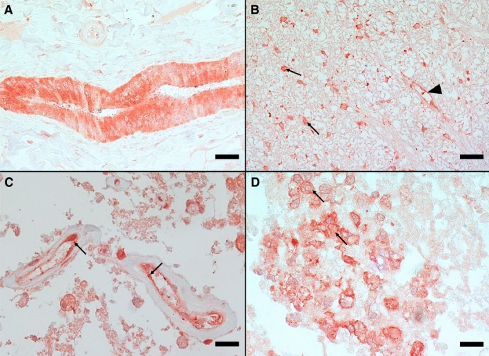
Figure 3.

Immunohistochemistry for endothelin‐1 of canine liver (A) and spinal cord (B–D) tissue. A—Marked immunostaining of bile duct in liver serving as positive control (IHC, bar = 50 μm); B—Spinal cord white matter with strongly stained astrocytes (arrows) and a small capillary with positively stained endothelial cells (arrowhead) (IHC, bar = 50 μm); C—Severely necrotic area in the center of the lesion with small vessels with positively stained endothelial cells (arrows) (IHC, bar = 20 μm); and D—Positively stained macrophages (arrows) (IHC, bar = 20 μm).
