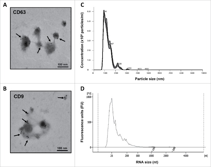
Figure 1.
Morphological characterization of breastmilk EVs and size distribution of their RNA cargo. (A) EVs immune-gold labeled by anti-CD63 antibody. (B) EVs immune-gold labeled by anti-CD9 antibody. (C) Concentration and size distribution of EVs in breastmilk by nanoparticle tracking analysis using the NanoSight NS300. (D) Size distribution of EV-encapsulated RNA showing the presence of lncRNAs, as measured by Agilent 2100 Bioanalyzer. Transmission electron microscopy images for (A) and (B) were taken by a JEOL 1200EX microscope. Arrows indicate positive staining.

