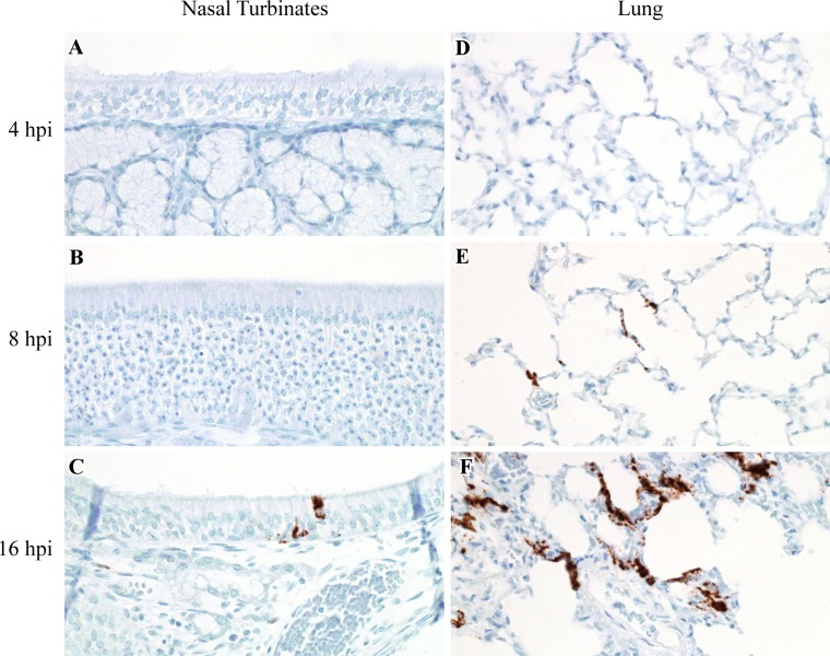
Fig 2. Virus replication in the nasal cavity and lung in hamsters inoculated with NiV-M.
ISH was used to detect positive sense viral RNA, indicating virus replication, in the nasal cavity and lung at 4, 8 and 16 hpi in Syrian hamsters intranasally inoculated with NiV-M. Viral RNA is labeled brown in all images. Virus replication was not present in the nasal cavity at 4 or 8 hpi (A, B), but was observed at 16 hpi, as shown here in the respiratory epithelium lining a nasal turbinate (C). Virus replication was not identified at 4 hpi in the lung (D), yet was detected in pneumocytes at 8 and 16 hpi (E, F). All images were taken at 400x. A schematic representation of the hamster respiratory tract is shown in Fig 5.

