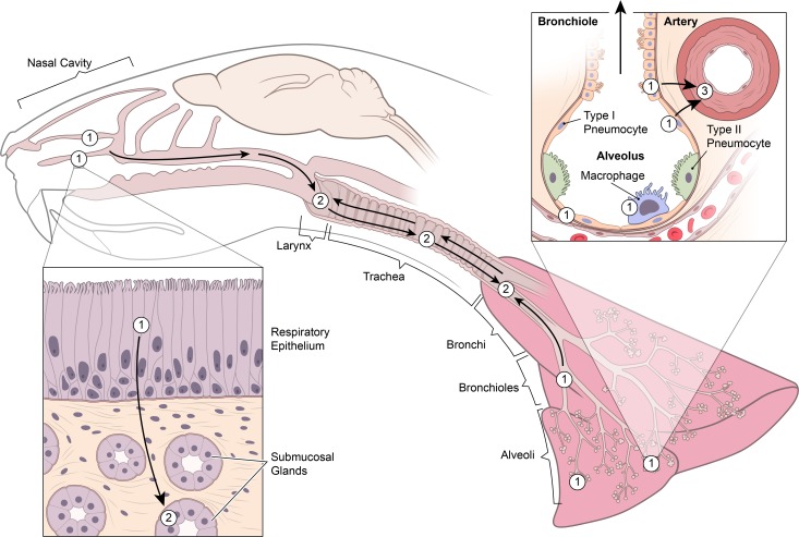
Fig 5. Schematic representation of the early dissemination of NiV through the upper and lower respiratory tract.
Nipah virus initially infects respiratory and olfactory epithelium lining the nasal turbinates and type I pneumocytes, bronchiolar respiratory epithelium and alveolar macrophages in the lung (1). Nipah virus then spreads from the epithelium in the nasal cavity to the underlying submucosal glands (2). At the same time, the virus also moves downward from the nasal cavity and/or upward from the lung to infect epithelial cells lining the larynx, trachea and bronchi (2). Subsequently, Nipah virus infects the smooth muscle of arterial walls in the lung, likely as a result of local spread from adjacent infected pneumocytes and bronchiolar respiratory epithelial cells (3). Of note, NiV-M and NiV-B exhibit a similar dissemination pattern; however, the rate of spread is slower with NiV-B.

