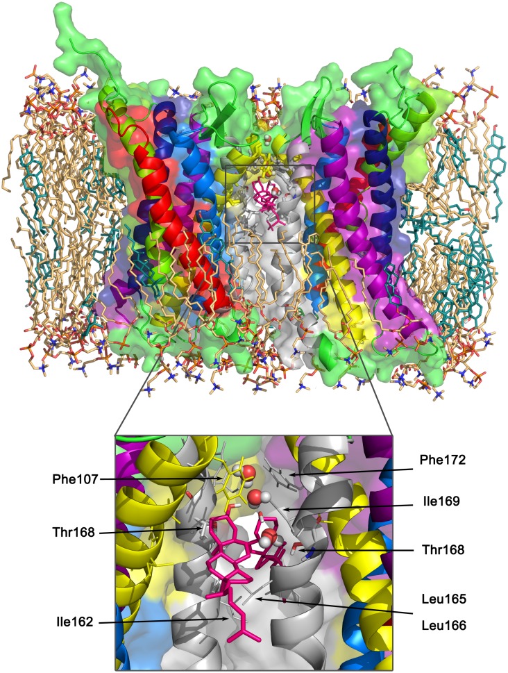
Fig 5. Cholesterol intercalation at the cholesterol-induced dimeric interface.
TM3,4/TM3,4 dimer F with two symmetrically intercalated cholesterols (shown as hotpink sticks) (structure after 100 ns atomistic MD simulation). The surrounding membrane is shown in light orange (POPC) and teal (cholesterol) sticks. In the insert, the two intercalating cholesterol molecules are highlighted together with their surroundings and water molecules (shown as spheres) forming bridges between the cholesterols and between Thr168 and cholesterol.

