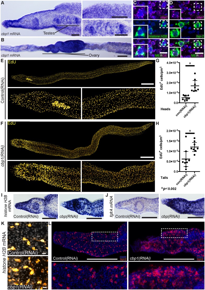
Fig 1. cbp1(RNAi) results in an increase in neoblast proliferation.
(A, B) Whole mount in situ hybridization showing cbp1 mRNA expression in (A) male and (B) female parasites. cbp1 is expressed broadly in the worm including in the testes and ovaries. (C, D) FISH showing expression of cbp1 with (C) Histone H2B and (D) tsp-2. Nuclei are in blue. (E, F) EdU labeling showing cell proliferation in (E) control and (F) cbp1(RNAi) male parasites. Images are tiled from multiple confocal stacks acquired from parasites at D14 of RNAi. Parasites were fixed after an overnight EdU pulse. (G,H) Quantification of EdU+ neoblasts per μm3 from (G) heads and (H) tails. Each dot represents counts from a confocal stack taken from a single male parasite and error bars represent 95% confidence intervals. Differences are statistically significant (p < 0.002, t-test). Quantification was performed on male parasites at D14 of RNAi. (I,J) WISH for neoblast markers (I) Histone H2B and (J) fgfrA in control and cbp1(RNAi) animals. Images are from D14 of RNAi and are representative of n > 10 male parasites. (K) FISH for Histone H2B in control and cbp1(RNAi) animals. Nuclei are in gray. Images are from D11 of RNAi. (L) FISH for tsp-2 in control and cbp1(RNAi) animals. D13 of RNAi. Anterior faces left in A-B, E-F, I, J, and L. Scale Bars: A,B 100 μm; C,D, and K 10 μm; E,F 500 μm; I,J, and L 250 μm.

