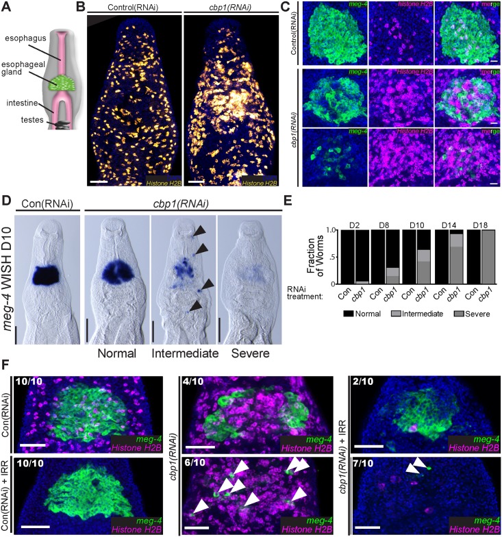
Fig 2. cbp1(RNAi) results in degeneration of the esophageal glands.
(A) Cartoon depicting the position of the schistosome esophageal gland relative to other tissues. (B) FISH for Histone H2B showing a mass of proliferative neoblasts in proximity to the esophageal glands at D11 of RNAi treatment. (C) Double FISH for the esophageal gland marker meg-4 and the neoblast marker Histone H2B following 11 days of control or cbp1 RNAi treatments. The number of esophageal gland cells is reduced and the number of proliferative neoblasts in increased. In extreme cases only a few meg-4 + cells remain in cbp1(RNAi) animals. Top and bottom panels for cbp1(RNAi) depict animals with differing phenotypic severities. Nuclei are in blue. (D) WISH for meg-4 at D10 of RNAi treatment. At this time point, by WISH three distinct phenotypic severities are observed: normal, intermediate, and severe. Normal animals possess relatively normal gland structure and meg-4 + labeling. Intermediate animals have clearly abnormal or degenerated gland structure and often possess ectopic meg-4 + cells (arrowhead) outside of the region were the gland resides in control animals. Severe animals possess few, if any, meg-4 + cells. (E) Plots depicting the relative fraction of animals that display normal, intermediate, or severe phenotypes with respect to the esophageal glands as observed by WISH for meg-4 at D2–D18 of RNAi. >12 animals from two separate experiments were observed for each time point. (F) Double FISH for Histone H2B and meg-4 at D11 in irradiated (+IRR) and unirradiated worms. Degeneration of esophageal glands in cbp1(RNAi) worms is not abrogated following irradiation. However, notably more isolated meg-4 + cells (Arrowheads) remain in unirradiated cbp1(RNAi) worms. Numbers indicate fraction of worms displaying phenotypes similar to those pictured. Nuclei are in blue. Anterior faces up in all images. Scale Bars: B, F 50 μm; C 20 μm; D 100 μm.

