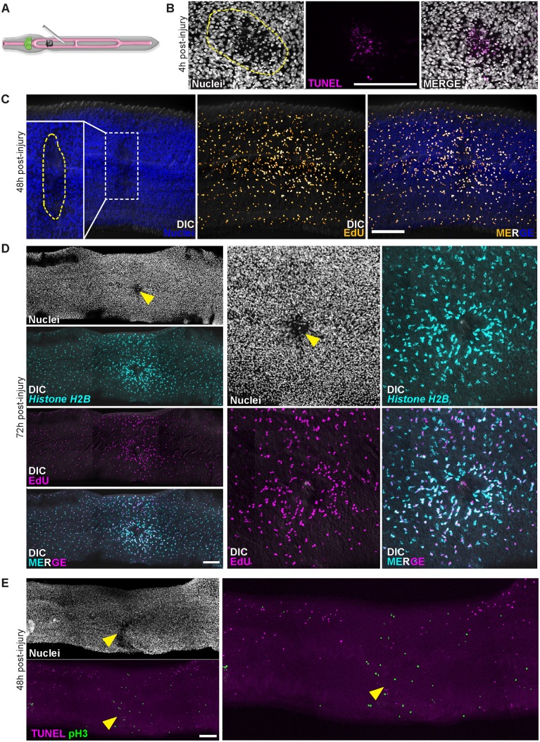
Fig 4. Physical injury initially induces cell death and increases in neoblast proliferation.
(A) Cartoon showing strategy to injure worms. (B) 4 hours post-injury the number of TUNEL+ cells is increased at the site of injury. Dashed line approximates the site of injury. Images representative of n = 21/23 male parasites. (C) EdU-incorporating neoblasts accumulate near wound site at 48 hours post-injury (n = 18/19 parasites). Parasites were fixed after a 4 hour EdU pulse. Dashed line in inset approximates the site of injury. (D) Histone H2B + neoblasts at 72 hours post-injury are enriched around wound sites (n = 11/11 male parasites). Parasites were labeled with EdU for 4 hours at 48 hours post-injury and were fixed 24 hours later (72 hours post-injury). Arrowhead indicates approximate site of injury. (E) TUNEL staining for cell death and Phospho-Histone H3 (pH3) labeling for neoblasts in mitosis in animals at 48 hours post-injury. Mitotic neoblasts 48 hours post-injury are clustered at wound sites (n = 28/29 parasites), whereas the number of TUNEL+ cells are reduced in tissues adjacent to wound sites (n = 24/26 male parasites). Arrowhead indicates approximate site of injury. Scale Bars: 100 μm. D and E are titled images from multiple confocal stacks.

