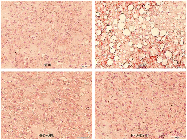
Fig 3. Effect of DSHT on HFD-induced histopathological changes of liver tissue stained with oil red O staining.
(A) Representative image of liver tissue of normal control animal (NOR) stained with Oil red O. (B) Representative image of liver tissue of animals treated with HFD. (C) Representative image of liver tissue of animals treated HFD+ORL. (D) Representative image of liver tissue of HFD+DSHT treated animals. Pathophysiological examination of the tissue sections was performed under light microscopy with 200x magnification.

