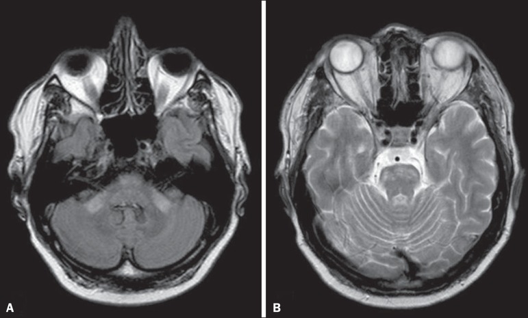
Figure 5.
Neuro-Behçet's disease. Axial FLAIR MRI sequence (A) and axial T2-weighted MRI sequence (B) showing, at the base of the cerebral peduncle, heterogeneous lesion bilaterally at the mesodiencephalic junction with extensive swelling, sparing the red nucleus. Note the cranial extent of the perilesional edema.

